Changing stroke rehab and research worldwide now.Time is Brain! trillions and trillions of neurons that DIE each day because there are NO effective hyperacute therapies besides tPA(only 12% effective). I have 523 posts on hyperacute therapy, enough for researchers to spend decades proving them out. These are my personal ideas and blog on stroke rehabilitation and stroke research. Do not attempt any of these without checking with your medical provider. Unless you join me in agitating, when you need these therapies they won't be there.

What this blog is for:

My blog is not to help survivors recover, it is to have the 10 million yearly stroke survivors light fires underneath their doctors, stroke hospitals and stroke researchers to get stroke solved. 100% recovery. The stroke medical world is completely failing at that goal, they don't even have it as a goal. Shortly after getting out of the hospital and getting NO information on the process or protocols of stroke rehabilitation and recovery I started searching on the internet and found that no other survivor received useful information. This is an attempt to cover all stroke rehabilitation information that should be readily available to survivors so they can talk with informed knowledge to their medical staff. It lays out what needs to be done to get stroke survivors closer to 100% recovery. It's quite disgusting that this information is not available from every stroke association and doctors group.

Friday, April 19, 2024

Treatment Relieves Chronic Lyme Disease’s Neurological Symptoms

With even the minutest functioning brain in stroke leadership, they should easily see that this research needs to be replicated in stroke survivors . But since there is NO leadership in stroke; NOTHING WILL BE DONE!

Treatment Relieves Chronic Lyme Disease’s Neurological Symptoms

Summary: Fibroblast growth factor receptor inhibitors, commonly used in cancer treatment, could effectively reduce neurological symptoms in patients with post-treatment Lyme disease syndrome. The study shows these inhibitors can decrease inflammation and cell death in brain and nerve tissues affected by Lyme disease.

This discovery paves the way for potential treatments aimed at the persistent neuroinflammation seen in some patients after standard antibiotic therapy. With promising initial results, further research is essential to move these findings from the lab to clinical applications.

Key Facts:

  1. Lyme disease can cause persistent neurological symptoms such as memory loss and fatigue, known as post-treatment Lyme disease syndrome, even after antibiotics.
  2. The study found that targeting FGFR pathways with specific inhibitors can significantly reduce inflammation and neuronal damage in tissue samples infected with Lyme disease bacteria.
  3. The research, supported by the Bay Area Lyme Foundation and resources from the Tulane National Primate Research Center, marks a critical step toward developing new interventions for chronic Lyme disease complications.

Source: Tulane University

Tulane University researchers have identified a promising new approach to treating persistent neurological symptoms associated with Lyme disease, offering hope to patients who suffer from long-term effects of the bacterial infection, even after antibiotic treatment.

Their results were published in Frontiers in Immunology

Lyme disease, caused by the bacterium Borrelia burgdorferi and transmitted through tick bites, can lead to a range of symptoms, including those affecting the central and peripheral nervous systems.

While antibiotics can effectively clear the infection in most cases, a subset of patients continues to experience symptoms such as memory loss, fatigue, and pain—a condition often referred to as post-treatment Lyme disease syndrome. 

Principal investigator Geetha Parthasarathy, PhD, an assistant professor of microbiology and immunology at the Tulane National Primate Research Center, has discovered that fibroblast growth factor receptor inhibitors, a type of drug previously studied in the context of cancer, can significantly reduce inflammation and cell death in brain and nerve tissue samples infected with Borrelia burgdorferi.

This discovery suggests that targeting FGFR pathways may offer an exciting new therapeutic approach to addressing persistent neuroinflammation in patients with post-treatment Lyme disease syndrome.  

“Our findings open the door to new research approaches that can help us support patients suffering from the lasting effects of Lyme disease,” Parthasarathy said.

“By focusing on the underlying inflammation that contributes to these symptoms, we hope to develop treatments that can improve the quality of life for those affected by this debilitating condition.”

Researchers treated nerve tissue with live or inactivated Borrelia burgdorferi, followed by an application of FGFR inhibitors. Study results revealed a significant reduction in both inflammatory markers and of cell death. 

While further research is needed to translate these findings into clinical treatments, the study represents an important step forward in understanding and potentially managing the complex aftermath of Lyme disease.

Funding: This study was funded by the Bay Area Lyme Foundation and supported with resources from the Tulane National Primate Research Center base grant of the National Institutes of Health, P51 OD011104. 

About this Lyme disease and neuropharmacology research news

Author: Keith Brannon
Source: Tulane University
Contact: Keith Brannon – Tulane University
Image: The image is credited to Neuroscience News

Original Research: Open access.

Upper limb motor recovery post-stroke seminar: Embracing complexity to deliver optimal outcomes.

When your doctors and therapists go to this event; DEMAND THEY COME BACK WITH 100% RECOVERY PROTOCOLS! Anything less is a waste of time.

 Upper limb motor recovery post-stroke seminar: Embracing complexity to deliver optimal outcomes.

Event Location

33 Queen Square Basement Lecture Theatre
Location
33 Queen Square Basement Lecture Theatre
Venue Details

Basement Lecture Theatre

33 Queen Square

London

WC1N 3BG

More Information

How to get to Queen Square 

Arriving by national rail :

Buses leave from the following mainline train stations to Russell Square:

Euston - 68, 168, 59, 91

King's Cross - 35, 91

Waterloo - 59, 168, 188

Charing Cross - 91

Paddington - 7 

Arriving by car:

Parking is available at the NCP Brunswick Square car park on Marchmont Street, LondonWC1N 1AF, which is a five minute walk to Guildford Street.

MAP


Arriving by tube: 

The nearest tube stations are Russell Square, Euston and King’s Cross.

Please click on the link below for a map of the area:

TUBE

 
Date of Event
13th May 2024
Last Booking Date for this Event
13th May 2024

Description

Upper limb motor recovery post-stroke: Embracing complexity to deliver optimal outcomes.

Dr Kate Hayward, University of Melbourne


Dr Kate Hayward is an Associate Professor of Stroke Recovery and Rehabilitation at the University of Melbourne. Kate is an NHMRC Emerging Leader, Heart Foundation Future Leader, and Dame Kate Campbell Fellow. She leads the REPAIR Research Group which applies early phase clinical trial designs to study when the ideal time is to start therapy after stroke, how much therapy should be provided, and who stands to benefit most from therapy. Kate also co-Chaired the recent third international Stroke Recovery and Rehabilitation Roundtable.


The seminar will take place in person with no online attendance option. Please join us after the event for wine, soft drinks, crisps, and networking. 

Attendee CategoryCostPlace(s) Available  

Free.£0.0052[Read More]


Thursday, April 18, 2024

Obesity-induced cognitive decline: Role of brain oxidation and tocotrienols

Your doctor has had a long time to come up with a protocol for tocotrienols. Did they? Or were they incompetent in this also like they were in everything else about stroke recovery? Did they get human testing initiated?


Obesity-induced cognitive decline: Role of brain oxidation and tocotrienols

Health Topics mentioned in this article Obesity
Alzheimers Disease
Cognitive Impairment Researchers from Shibaura Institute of Technology, Japan investigated the impact of HFSD on mice and found that tocotrienols (T3s), a form of vitamin E, show promise in protecting against cognitive decline associated with obesity, highlighting its therapeutic potential.
Obesity has become a pressing worldwide health issue, with rates steadily rising over recent decades. Beyond its well-documented associations with physical health issues, such as cardiovascular disease and diabetes, obesity has also been linked to cognitive decline, including conditions like Alzheimer's disease and Parkinson's disease. Understanding the complex mechanisms underlying this cognitive impairment is crucial for developing effective interventions. In a study published in the International Journal of Molecular Sciences, Assistant Professor Yugo Kato from Tottori University and Shibaura Institute of Technology, Professor Koji Fukui from Shibaura Institute of Technology and their team offer insights into potential solutions.The study investigated the neuroprotective effects of tocotrienols (T3s) in mitigating the adverse impact of diet-induced obesity on brain function."Our goal is to combat obesity-related diseases using natural compounds and thereby reduce the prevalence of conditions like dementia among individuals affected by obesity," says Prof. Kato.

T3s are a group of naturally occurring chemical compounds belonging to the vitamin E family. Past studies have revealed that T3s have neuroprotective and anti-obesity properties. Additionally, they have also been shown to pass through the blood-brain barrier and enter cells to produce antioxidant effects. However, little is known about how T3s contribute to the decline in brain function brought on by obesity.

Quality Settings 12 Years of Data Link Popular Diet to Brain BoostHigh Adolescent BMI Linked With Chronic Kidney Disease, Study Warns To address this gap, the team conducted a comprehensive investigation using a mouse model system. They employed a meticulous experimental design, with C57BL/6 male mice subjected to either a high-fat, high-sucrose diet (HFSD) or a control diet, supplemented with or without T3s. In the initial phase of the study, the team evaluated the anti-obesity effects of T3s. To do so, they incorporated a 50mg T3s mixture into 100g of both experimental diets, namely the control and HFSD. Key parameters, including body weight, fat deposition, serum cholesterol, triglyceride, and glucose concentrations, were assessed alongside cognitive function using the Morris water maze and Y-maze tests. Additionally, markers of oxidative stress and proteomic changes in the cortex were analyzed to gain deeper insights into the underlying mechanisms.The results of the study were highly promising. While HFSD feeding induced obesity in the mice, supplementation with T3s did not mitigate weight gain. However, T3s treatment demonstrated a significant improvement in cognitive function, as evidenced by enhanced learning ability in HFSD-fed mice. Furthermore, the study revealed the role of oxidative stress in obesity-induced cognitive decline, with HFSD-fed mice exhibiting increased brain oxidation levels. Remarkably, T3s treatment appeared to mitigate this oxidative stress, suggesting a potential mechanism for their neuroprotective effects. Additionally, contrary to expectations, the team found that respiratory metabolism decreased and the temperature around brown adipose tissue increased in mice fed with HFSD. This unexpected finding suggests that HFSD may have a complex impact on metabolic processes and temperature regulation in the body."Lastly, we wanted to examine the protein change associated with the consumption of the HFSD. So, we performed a quantitative proteomic analysis of the mouse cortex. Our focus was on the proteins that were expressed differently between the HFSD and control groups," explains Prof. Kato. They discovered that in comparison to the control group, obesity brought on by HFSD feeding changed 12 proteins, and mice treated with T3s showed considerable prevention of these changes. In conclusion, this study represents a significant step forward in our understanding of the intricate relationship between obesity and cognitive decline. By uncovering the potential benefits of T3s in preserving cognitive function, the research opens up new avenues for therapeutic interventions targeting neurodegenerative diseases associated with obesity.More information: Yugo Kato et al, Tocotrienols Prevent the Decline of Learning Ability in High-Fat, High-Sucrose Diet-Fed C57BL/6 Mice, International Journal of Molecular Sciences (2024). DOI: 10.3390/ijms25063561Provided by Shibaura Institute of TechnologyThis story was originally published on Medical Xpress. Subscribe to our newsletter for the latest sci-tech news

Direction-specific Disruption of Paretic Arm Movement in Post-stroke Patients

 I'm missing how anything here is going to get survivors recovered!

Direction-specific Disruption of Paretic Arm Movement in Post-stroke Patients

Kiyoshi Yoshioka, RPT, PhD a,* Tatsunori Watanabe, RPT, PhD b,* Mizuki Yoshioka, RPT a Keita Iino, RPT a Kimikazu Honda, RPT a Koshiro Hayashida, RPT a and Yuji Kuninaka, RPT a
Received: January 17, 2020, Accepted: April 8, 2020, Published online: April 17, 2020 
 a Kumamoto Center, Rehabilitation Center for all Customers with Stroke and Cerebrovascular Diseases, SENSTYLE Inc., Kumamoto, Japan  
b Department of Sensorimotor Neuroscience, Graduate School of Biomedical and Health Sciences, Hiroshima University, Hiroshima, Japan Correspondence: Kiyoshi Yoshioka, RPT, PhD, Institute for Research on Productive Aging (IRPA), #201 Kobe Hybrid Business Center, Minami-cho 6-7-6, Minatojima, Kobe 650-0047, Japan, Email: kiyoshi.yoshioka.y@gmail.com 
 
Objective:  
 
This study aimed to characterize reaching movements of the paretic arm in diferent directions within the reachable workspace in post-stroke patients.  
 
Methods:  
 
A total of 12 post- stroke patients participated in this study. Each held a ball with a tracking marker and performed back-and-forth reaching movements from near the middle of the body to one of two targets in front of them located on the ipsilateral and contralateral sides of the arm performing the movement. We recorded and analyzed the trajectories of the tracking marker. The stability of arm movements was evaluated using areas and minimum Feret diameters to assess the trajectories of both the paretic and non-paretic arms. The speed of the arm movement was also calculated.  
 
Results:  
 
For the paretic arm, contralateral movement was more impaired than ipsilateral movement, whereas for the non-paretic arm, no diference was observed between the directions. The maximum speed of the contralateral movement was signifcantly slower than that of the ipsilateral movement in both the paretic and non-paretic arms.  
 
Conclusion:  
 
The paretic arm shows direction specifc instability in movement toward the contralateral side of the arm.

Thought Provoking Work May Reduce Later Life Cognitive Decline

 My last six years of working were as a mainframe computer programmer for IBM as a consultant, so I got dumped into problem situations with no systems employees still there(just offshore contractors) and no one on the business side knew what the systems were supposed to do. And we were expected to solve the problems in weeks, not months.

Thought Provoking Work May Reduce Later Life Cognitive Decline

Summary: Engaging in complex, thought-provoking work may lower the risk of mild cognitive impairment (MCI) in older age. Conducted by researchers at Oslo University Hospital, the study analyzed job demands of over 7,000 people across 305 occupations in Norway, linking higher cognitive job demands in one’s 30s through 60s to a reduced incidence of MCI post-70.

The research categorized jobs by the nature of tasks—routine manual, routine cognitive, non-routine analytical, and non-routine interpersonal—with teaching, a high cognitive demand job, showing lower rates of MCI compared to less demanding roles like mail carrying. Adjustments for demographic and lifestyle factors still showed a 66% higher risk of MCI in those with the least cognitively demanding jobs.

Key Facts:

  1. The study involved an extensive demographic, assessing cognitive job demands and their impact on 7,000 individuals aged 70 and above.
  2. Participants in the highest cognitive demand jobs had a significantly lower rate of mild cognitive impairment (27%) compared to those in the lowest demand jobs (42%).
  3. The research supports the notion that intellectually stimulating work can serve as a protective factor against cognitive decline later in life, although further studies are needed to identify specific beneficial tasks.

Source: AAN

The harder your brain works at your job, the less likely you may be to have memory and thinking problems later in life, according to a new study published in the April 17, 2024, online issue of Neurology.

This study does not prove that stimulating work prevents mild cognitive impairment. It only shows an association.

“We examined the demands of various jobs and found that cognitive stimulation at work during different stages in life—during your 30s, 40s, 50s and 60s—was linked to a reduced risk of mild cognitive impairment after the age of 70,” said study author Trine Holt Edwin, MD, PhD, of Oslo University Hospital in Norway.

“Our findings highlight the value of having a job that requires more complex thinking as a way to possibly maintain memory and thinking in old age.”

The study looked at 7,000 people and 305 occupations in Norway.

Researchers measured the degree of cognitive stimulation that participants experienced while on the job. They measured the degree of routine manual, routine cognitive, non-routine analytical, and non-routine interpersonal tasks, which are skill sets that different jobs demand.

Routine manual tasks demand speed, control over equipment, and often involve repetitive motions, typical of factory work. Routine cognitive tasks demand precision and accuracy of repetitive tasks, such as in bookkeeping and filing.

Non-routine analytical tasks refer to activities that involve analyzing information, engaging in creative thinking and interpreting information for others. Non-routine interpersonal tasks refer to establishing and maintaining personal relationships, motivating others and coaching. Non-routine cognitive jobs include public relations and computer programing.

Researchers divided participants into four groups based on the degree of cognitive stimulation that they experienced in their jobs.

The most common job for the group with the highest cognitive demands was teaching. The most common jobs for the group with the lowest cognitive demands were mail carriers(My dad was one and ended up with Parkinsons and dementia) and custodians.

After age 70, participants completed memory and thinking tests to assess whether they had mild cognitive impairment. Of those with the lowest cognitive demands, 42% were diagnosed with mild cognitive impairment. Of those with the highest cognitive demands, 27% were diagnosed with mild cognitive impairment.

After adjustment for age, sex, education, income and lifestyle factors, the group with the lowest cognitive demands at work had a 66% higher risk of mild cognitive impairment compared to the group with the highest cognitive demands at work.  

“These results indicate that both education and doing work that challenges your brain during your career play a crucial role in lowering the risk of cognitive impairment later in life,” Edwin said.

“Further research is required to pinpoint the specific cognitively challenging occupational tasks that are most beneficial for maintaining thinking and memory skills.”

A limitation of the study was that even within identical job titles, individuals might perform different tasks and experience different cognitive demands.

Funding: The study is supported by the National Institutes of Health.

About this cognition and aging research news

Author: Natalie Conrad
Source: AAN
Contact: Natalie Conrad – AAN
Image: The image is credited to Neuroscience News
Study: Heart failure, not stroke, most common complication of atrial fibrillation

Interesting.


Study: Heart failure, not stroke, most common complication of atrial fibrillation

The threat of heart failure from atrial fibrillation could be more worrisome than stroke, a new study reveals. Photo by mirkosajkov/Pixabay
1 of 2 | The threat of heart failure from atrial fibrillation could be more worrisome than stroke, a new study reveals. Photo by mirkosajkov/Pixabay

NEW YORK, April 17 (UPI) -- Doctors often tell patients with atrial fibrillation, which causes an irregular and often abnormally rapid heart rate, to beware of an increased risk of stroke. But the threat of heart failure should be even more worrisome, a new study reveals.

The study, conducted at Aalborg University in Denmark in collaboration with Tufts Medical Center and Boston Medical Center, was published Wednesday in the British Medical Journal.

"Our nationwide study shows that the lifetime risk of atrial fibrillation increased over the past two decades from one in four to one in three," the authors wrote.

"After atrial fibrillation, heart failure was the most frequent complication, with a lifetime risk of two in five, twice greater than the lifetime risk of stroke after atrial fibrillation."

Even so, more research is needed to find new and effective preventive strategies to further decrease stroke and heart failure risks, the authors noted. They called for alignment of both randomized, clinical trials and guidelines "to better reflect the needs of the real-world population with atrial fibrillation."

Atrial fibrillation will affect 16 million people in the United States by 2050 and 18 million people in Europe by 2060, according to the researchers, who received a grant from the Danish Cardiovascular Academy to undertake the study.

"Atrial fibrillation is a very common heart rhythm disorder," the study's lead author, Dr. Nicklas Vinter, a postdoctoral researcher in the department of clinical medicine at Aalborg University, told UPI via email.

"We wanted to understand the long-term risks better and see how these risks have changed over time," Vinter said, adding that "in many cases, living a healthy lifestyle and taking your prescribed medicine can prevent atrial fibrillation and its complications."

National data analyzed

To address a knowledge gap, researchers analyzed national data for 3.5 million Danish adults with no history of atrial fibrillation at age 45 or older to know whether they developed the condition over a 23-year period spanning 2000 to 2022.

The researchers subsequently followed all 362,721 individuals with a new diagnosis of atrial fibrillation (46% women and 54% men), but without complications, until they had a diagnosis of heart failure, stroke or heart attack.

They took into account potentially influential factors, such as history of high blood pressure, diabetes, high cholesterol, heart failure, chronic lung and kidney disease, family income and educational attainment.

The results indicated that the lifetime risk of atrial fibrillation increased to 31% om 2011-12 from 24% in 2000-2010. The increase was greater among men and individuals with a history of heart failure, heart attack, stroke, diabetes and chronic kidney disease.

Among those with atrial fibrillation, the most common complication was heart failure (lifetime risk 41%). This was twice as large as the lifetime risk of any stroke (21%) and four times greater than the lifetime risk of heart attack (12%).

Men showed a higher lifetime risk of complications after atrial fibrillation compared with women for heart failure (44% versus 35%) and heart attack (12% versus 10%), while the lifetime risk of stroke after atrial fibrillation was slightly lower in men than women (21% vs 23%).

Over the 23-year study period, there was virtually no improvement in the lifetime risk of heart failure after atrial fibrillation (43% in 2000-10 vs 42% in 2011-22) and only slight (4% to 5%) decreases in the lifetime risks of any stroke, ischemic stroke and heart attack after atrial fibrillation, which were similar among men and women.

Among those with atrial fibrillation, 2 in 5 most likely will develop heart failure and 1 in 5 will encounter a stroke during their remaining lifetime, with little or no improvement in risk evident over the 20-year study period, the study indicated.

Strategies needed

As a result, the researchers pointed out that strategies to prevent both heart failure and stroke are necessary for people with atrial fibrillation.

However, this was an observational study, so researchers could not draw firm conclusions about cause and effect, and they acknowledged potentially missing patients with undiagnosed atrial fibrillation.

They also lacked information about ethnicity or lifestyle factors, and the results may not apply to other countries or settings.

"This study is in line with prior published data from the U.S. that shows an increasing prevalence of atrial fibrillation," said Dr. Timothy Larsen, a cardiologist and cardiac electrophysiologist at Rush University System for Health in Chicago. He was not involved in the research.

While heart palpitations are a classic symptom of atrial fibrillation, patients also should pay attention to more subtle or generalized symptoms, such as fatigue and shortness of breath, which usually occurs with exertion. Controlling atrial fibrillation can improve these symptoms, Larsen said.

Many patients have been able to prevent stroke and death --"the most devastating consequences" of atrial fibrillation -- by taking very safe and effective drugs, such as blood thinners, said Dr. Gregory Marcus, a cardiologist and endowed professor of atrial fibrillation research at the University of California-San Francisco.

As a result, "it's important to emphasize that heart failure and not stroke is the most common complication of atrial fibrillation specifically in this modern era," Marcus said.

Strong evidence demonstrates that "eradication of atrial fibrillation with catheter ablation procedures can substantially improve heart failure and even significantly prolong longevity," he added.

Despite the new emphasis on heart failure as a complication of atrial fibrillation, the risk of stroke can't be understated, said Dr. Paul Johnson, medical director of the Comprehensive Stroke Center at Intermountain Medical Center in Murray, Utah.

As a neurohospitalist and vascular neurologist, Johnson said he recommends that patients remember the acronym BEFAST, as suggested by many health systems, to remain alert for signs of a stroke.

The first five letters stand for sudden changes in balance, eyes, face, arms and speech, while the "T" represents time because calling 911 immediately is critical to prevent brain damage.

Both the American Stroke Association and the Centers for Disease Control and Prevention abbreviate the acronym to FAST because acting fast is key to stroke survival.

"This study draws attention to the high prevalence of atrial fibrillation in the general population, and in fact, suggests that the prevalence of this condition is increasing," Johnson said.

"Because atrial fibrillation is associated with several significant medical complications, including stroke and heart failure, this represents a serious public health issue."

Georgia Coverdell Acute Stroke Registry

 Look how appalling this is! Improving 'care', NOT RESULTS OR RECOVERY! This is why we need survivors in charge; we would single mindedly focus on 100% recovery; NOT THIS USELESS 'CARE' CRAPOLA!

Send me hate mail on this: oc1dean@gmail.com. I'll print your complete statement with name and my response in my blog. Or are you afraid to engage with my stroke-addled mind?  Survivors would like to know why you are doing useless stuff.

Georgia Coverdell Acute Stroke Registry

PROGRAM HISTORY

Named in honor of the late Senator Paul Coverdell of Georgia, who died of a massive stroke in 2000, the primary goal of the Georgia Coverdell Acute Stroke Registry (GCASR) program is to improve the care of acute stroke patients in the hospital and pre-hospital settings. The program is funded by the Centers for Disease Control Paul S. Coverdell National Acute Stroke Registry cooperative agreement and was established in 2001 as a prototype project implemented by the Emory University School of Medicine. Full implementation and incorporation into the State’s Department of Public Health (DPH) began in 2005.       

PURPOSE

This program addresses quality improvement in multiple areas of stroke care, from rapid screening, diagnosis, and intervention for patients experiencing an acute stroke, to secondary prevention measures such as blood pressure control, smoking cessation, and treatment of elevated cholesterol to reduce the incidence of recurrent stroke after hospital discharge. In addition, the program will also help improve the use of rehabilitation services for those who have experienced an acute stroke, in an effort to reduce long-term disability due to stroke.

States in the southeastern U.S. have the highest incidence and mortality of stroke; as a result, this area of the U.S. is commonly referred to as the stroke belt. In an effort to reduce long-term disability, the GCASR and the State of Georgia Cardiovascular Health Initiative Program work to reduce heart disease-and stroke-related morbidity and mortality in Georgia.

View the following videos:

Coverdell Stroke Program: Ensuring that All Americans Receive the Highest-Quality Care 

Dr. Frankel’s Coverdell Story: Improving Stroke Care in Georgia 

GOALS

Increase Quality Improvement through collaborative efforts among participant hospitals, EMS agencies and Rehabilitation facilities

Lower the stroke morbidity experienced in Georgia.

Enhance the effectiveness of secondary care and prevent recurrent strokes.

Develop protocols to guide clinical care with effective stroke management.

Develop effective methods to care for acute stroke patients.

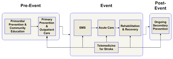
The Components of Stroke System of Care

Components of Stroke System of Care

PARTICIPATING HOSPITALS


F82 The 11th Annual Upper Limb Neurorehabilitation Course - London

 When your doctor or therapists go to this course, have them ask for 100% RECOVERY PROTOCOLS, not fucking useless guidelines and suggestions! Effective is 100% recovery! DO NOT ACCEPT ANYTHING LESS!

F82 The 11th Annual Upper Limb Neurorehabilitation Course - London

Event Information

QSUL

Location
33 Queen Square Basement Lecture Theatre
Venue Details

Basement Lecture Theatre

33 Queen Square

London

WC1N 3BG

 
Dates of Event
27th June 2024 – 28th June 2024
Last Booking Date for this Event
26th June 2024

Description

UCL Centre for Neurorehabilitation and the National Hospital for Neurology and Neurosurgery present:  

The 11th Annual Upper Limb Neurorehabilitation Course

Treating patients with upper limb deficit: Integrating research into practice 

Thursday 27th & Friday 28th June 2024.


This two-day course provides an up-to-date overview of current research in treatment and rehabilitation options for the neurological patient with upper limb deficit. The course looks at the practical, real-life translation of scientific evidence into clinical practice and discusses the ingredients that make an upper limb therapy effective. Delegates will have the opportunity to trial novel devices and robotic technology.


Enquiries to: cnr@ucl.ac.uk 


Attendee CategoryCost   

Standard Rate.£260.00[Read More]

Student Rate.£100.00[Read More

Long-term neuropsychiatric complications of aneurysmal subarachnoid hemorrhage: a narrative review

What are the EXACT PROTOCOLS your doctor is doing to prevent these occurrences? NONE? So you don't have a functioning stroke doctor, do you?

Post stroke depression(33% chance)

Post stroke anxiety(20% chance).  

Sexual dysfunction in stroke patients 1986

Long-term neuropsychiatric complications of aneurysmal subarachnoid hemorrhage: a narrative review

  1. Jose Danilo Bengzon Diestro1,2,
  2. Manav Vyas1,2,
  3. Youngkyung Jung3,
  4. Teruko Kishibe2,4,
  5. Carl Leochico1,5,6,7,
  6. Adrian Espiritu1,5,8,9,
  7. Maria Kristina Dorotan10,
  8. Nico Dimal1,
  9. Abdelsimar Tan Omar11,
  10. April Sienes12,
  11. Gustavo Saposnik1,2,
  12. Thomas R Marotta13,
  13. Atif Zafar1,
  14. Vitor Mendes Pereira3,
  15. Julian Spears3
  1. Correspondence to Dr Jose Danilo Bengzon Diestro, Department of Medicine, University of Toronto, Toronto, Canada; danni.diestro@gmail.com

Abstract

This review focuses on the often-neglected long-term neuropsychiatric consequences of aneurysmal subarachnoid hemorrhage (aSAH), beyond traditional randomized trial outcomes of mortality and retreatment. While current guidelines recommend screening for these sequalae, it may not be routinely practiced. This review will underscore the prevalence and management of common neuropsychiatric sequalae, including anxiety, depression, cognitive dysfunction, headaches, seizures, and sexual dysfunction, all of which can significantly impact the quality of life of survivors of aSAH. We emphasize the critical role neurointerventionalists can play by going beyond the customary practice of radiological monitoring for treated aneurysms by screening for and helping guide management of these common neuropsychiatric complications.


If you wish to reuse any or all of this article please use the link below which will take you to the Copyright Clearance Center’s RightsLink service. You will be able to get a quick price and instant permission to reuse the content in many different ways.

Background

Randomized trials examining the consequences of aneurysmal subarachnoid hemorrhage (aSAH) primarily concentrate on conventional outcomes, such as mortality, aneurysm recurrence, and aneurysm retreatment.1 2 While there is available data on functional outcomes assessed through the modified Rankin Scale (mRS), there is a paucity of studies dedicated to investigating long-term neuropsychiatric consequences, including anxiety, depression, epilepsy, chronic headaches, cognitive dysfunction, and sexual dysfunction. These outcomes can significantly impact patients' quality of life, even among those perceived to have favorable functional outcomes based on the mRS.3 4

Guidelines from the American Heart Association (AHA) recommend screening for these outcomes at discharge and during follow-up visits to facilitate timely intervention.5 As healthcare providers who routinely monitor treated aneurysms throughout patients' lifetimes, neurointerventionalists are well-situated to observe the progression and impact of these complications on patients’ quality of life. Our review aims to primarily elucidate the prevalence and management approaches for these long-term outcomes, offering practical screening tools suitable for the neurointerventional clinic setting. By doing so, we expect to enhance the long-term neuropsychiatric care of survivors of aSAH.

Method/search strategy

An information specialist (TK) worked with the lead author (JDBD) to develop the search strategy. We conducted the search on Medline (Ovid), Embase (Ovid), the Cochrane Central Register of Controlled Trials (Ovid), and Scopus (Elsevier). Keywords and Medical Subject Heading (MeSH) terms related to our research question were used. The search terms used included the combinations and truncations of the following: subarachnoid hemorrhage, mood disorders, depression, anxiety, seizure, epilepsy, convulsion, sexual dysfunction, dyspareunia, erectile dysfunction, impotence, vaginismus, premature ejaculation, cognition, mental fatigue, brain fog, neuropsychiatric, headache, cephalgia, and migraine. The full search strategy according to each database is available from the authors upon reasonable request.

Each outcome was addressed using a tailored search strategy for prevalence, and a separate distinct search strategy for questionnaires was employed across all outcomes. The results were then integrated into Covidence, an online systematic review tool aimed at enhancing collaboration and streamlining literature review processes. Lead authors (YJ, CL, AE, MKD, ND) were assigned to specific topics and provided access to a curated list of potential publications within their respective subtopics. Collaborating with the primary author (JDBD), they assessed the relevance and significance of these publications for inclusion in the review. Relevant articles were additionally manually searched to identify further pertinent literature.

Ethics

The study did not require ethics approval as it does not involve any patient data.

Anxiety and depression

Symptoms of depression and anxiety are relatively common in patients with intracranial aneurysms. Multiple evidence synthesis studies indicate that approximately three out of ten individuals who survived aSAH may develop symptoms of depression and anxiety (figure 1).6 7 Depression and anxiety symptoms associated with aSAH may have a substantial effect on patients’ lives. Systematic reviews revealed a negative association between depression/anxiety and health-related quality of life, and the presence of depression may also predict poorer quality of life in aSAH survivors.6 7 Depression was also found to be a significant predictor of unemployment.8 Depression after aSAH was found to be more common among females, those with premorbid depression or psychiatric illness, and those with substance use disorder. Further, having cognitive symptoms, fatigue, and higher physical disability also increases the risk of having depression after SAH.9

Figure 1

Summary of prevalence of long-term neuropsychiatic complications following aneurysmal subarachnoid hemorrhage (figure created with BioRender.com).

The 2023 American Heart Association (AHA)/American Stroke Association (ASA) guideline indicates that patients with aSAH with depression are recommended to undergo psychotherapy and drug therapy.5 Potential treatments for these symptoms were derived from therapies used for people with mental health conditions and from data on stroke survivors. Selective serotonin reuptake inhibitors (SSRIs) have been considered first-line therapy for major depressive and anxiety disorders.10 Among the SSRIs, fluoxetine has been recommended based on several randomized controlled trials (RCTs) in stroke patients.5 Furthermore, a recent 2023 Cochrane Living Systematic Review involving 65 trials on patients with stroke (n=5831) indicated significant reductions in reducing the prevalence of depression when they compared pharmacological therapies (SSRIs and other antidepressants) versus placebo (relative risk (RR) 0.70; 95% CI 0.55 to 0.88; 8 RCTs; n=1025), psychological (cognitive–behavioral therapy and others) versus usual care/attention control (RR 0.77; 95% CI 0.62 to 0.95; 6 trials; n=521), and the combination of non-invasive brain stimulation (transcranial magnetic stimulation) with pharmacological intervention versus pharmacological therapy only (RR 0.77; 95% CI 0.64 to 0.91; 3 RCTs; n=392).11

While improving outcomes in aSAH requires effective therapies for these psychiatric conditions, a prerequisite to this is the accurate identification of SAH survivors with anxiety and depressive disorders. A recent systematic review identified the nine most common depression and screening instruments used in patients with aSAH.12 These instruments include the Beck Depression Inventory-II (BDI-II), Hospital Anxiety and Depression Scale (HADS), Center for Epidemiologic Studies Depression Scale (CESDS), Zung Self-Rating Depression Scale, Geriatric Depression Scale, Montgomery–Asberg Depression Rating Scale (MADRS), Patient Health Questionnaire-9 (PHQ-9), EQ-5D Anxiety/Depression, and Stroke Specific Quality of Life. HADS has been validated for post-stroke patients and found to have a sensitivity of 86.8% and a specificity of 69.9% with a total cut-off score of 11.13 However, none of these tools have been validated for individuals with aSAH.12 Conducting further studies is essential to verify the validity of these screening techniques for aSAH.

Cognitive dysfunction

Cognitive impairment is a major sequela of aSAH seen in 40–70% of survivors and has been associated with more reduced quality of life measures (figure 1).14 It falls under the term post-stroke cognitive impairment, which refers to cognitive decline occurring 3 to 6 months after a stroke.15 Known risk factors for subsequent cognitive dysfunction following aSAH include acute hydrocephalus requiring cerebrospinal fluid diversion, seizures, fever, prolonged intensive care unit stay, and development of delayed cerebral ischemia, although even those with good functional outcomes and postoperative scores may still harbor cognitive deficits within the first 3 months to years later.14 16

The type of cognitive dysfunction depends on factors including location of the hemorrhage, artery affected, time to treatment, and comorbid factors,14 15 but domains more commonly involved include attention, executive function, and memory.14 16 Impairment in emotion recognition and social cognition can also be seen16 and may contribute to neuropsychiatric disturbances. Language deficits following aSAH remain understudied,14 although high performance on animal naming and abstraction, which are language-driven subtests, on cognitive testing like the Montreal Cognitive Assessment (MoCA) is more closely associated with returning to work following aSAH.17

All aSAH patients may undergo neurocognitive assessment for screening and longitudinal follow-up regardless of functional outcome.15 A proposed framework that captures the natural history of cognitive impairment and post-acute recovery involves a screening test done between onset up to 8–90 days later followed by in-depth testing done as early as 8–90 days up to 1 year and beyond if prior screening is positive.15 A recommended screening test is a 5-minute protocol including the Orientation, Memory, and Phonemic fluency MoCA subtests for assessment in the acute setting in addition to screening for delirium, which may impact longer-term cognitive outcomes.15 Following the acute phase, in-depth testing can be done using a validated global cognition screening test such as the full MoCA or the Mini-Mental State Examination (MMSE). The MoCA is an ideal and more sensitive screening tool than the MMSE as it affords a more granular assessment of executive, language, and visuospatial skills in addition to memory.17 Establishing cognitive function using these tools at 3 months post-aSAH is a useful measure and predictor of health-related quality of life (HRQoL) at 1-year follow-up.14

Once cognitive impairment is recognized, a holistic approach to rehabilitation may be instituted. It is found to be most beneficial around 6 months following aSAH.14 There remains, however, uncertainty as to the efficacy of cognitive rehabilitation interventions due to lack of robust data.15 Currently, there is no pharmacotherapy approved for post-stroke cognitive impairment.15

Headaches

While headaches are a prominent clinical feature of aSAH, their persistence after initial management and stabilization remains unclear. Reported rates of persistent headaches after aSAH vary between 32% and 41% in the literature, with some reporting severe headache years after the inciting event (figure 1).18 19 The natural history of post-aSAH headaches is also poorly defined, with no strict cut-off point after which SAH-related headaches should naturally subside. According to the International Classification of Headache Disorders (3rd edition), headaches persisting 3 months after resolution of aSAH are classified as “persistent headache attributed to past non-traumatic subarachnoid hemorrhage”.20

Unlike the classic thunderclap headache associated with the onset of aSAH, post-aSAH headaches tend to be more insidious in onset, often described as a sensation of pressure.18 21 There are no clear prognostic factors for developing chronic aSAH; clinical presentation, radiographic distribution, aneurysm type nor location, hydrocephalus, and management of the aSAH have not been associated with long-term headaches.18 19

In addition to knowing the type and character of the headaches, it is important to use questionnaires to understand their impact on a patient’s daily life. One center utilized the Headache Disability Index and a series of pain questionnaires (eg, the Short-Form McGill Pain Questionnaire (SF-MPQ), the German Pain Questionnaire, and the Depression, Anxiety Stress Scale (DASS)), along with the Health-Related Quality of Life (HRQOL) questionnaire and 12-item Short Form Health Survey.18 They found that higher headache scores correlated with reduced quality of life.

The management of post aSAH headaches is not well-defined, with no specific targeted therapies. In their narrative review of this topic, Sorrentino et al note the overall paucity of evidence in this area, with no evidence specifically focused on the outpatient management of post-aSAH headaches.21 Pharmaceutical management of headaches after aSAH heavily relies on a combination of acetaminophen, opioids, and steroids, with the vast majority of patients requiring more than one type of analgesic.22 Select studies also demonstrate a role for gabapentin, pregabalin, and intravenous lidocaine for the inpatient management of persistent headaches; however, the long-term effects are not well-known.21 Of note, most patients (95%) receive inpatient opioids and over 70% are discharged home with a prescription for opioids; however, there is little evidence that opioids improve headaches after the immediate SAH event.22

Seizures and epilepsy

Seizures and epilepsy are well-described complications of aSAH. Seizures are distinct from the diagnosis of epilepsy, which is characterized by an enduring predisposition for epileptic seizures.23 Seizures that occur within 24 hours to 14 days following ictus are considered acute symptomatic seizures which will likely not require long-term treatment, whereas those that occur >14 days are consistent with the diagnosis of epilepsy, requiring early diagnosis and long-term management.

An analysis of the ISAT study showed a higher risk of seizures for open surgery (9.6%) compared with endovascular therapy (5.2%) at 5 years.24 A population-based study showed a 5-year cumulative incidence of 12% (figure 1).25 Differences in reported incidence are likely related to inconsistencies between definitions of epilepsy, length of follow-up, confounding factors such as anti-seizure medications (ASM) prescribing practices, among others. Risk factors for developing epilepsy following aSAH include acute symptomatic seizures or seizures at onset, non-convulsive status epilepticus, middle cerebral artery aneurysms, higher Hunt and Hess and Fisher grades, and the presence of a large intracerebral hemorrhage.24 25

There is currently no gold standard or evidence-based guideline regarding the choice of ASM from epilepsy following aSAH. An earlier retrospective study of aSAH patients identified phenytoin exposure as a predictor of poor functional and cognitive outcomes.26 However, a subsequent observational study comparing levetiracetam to phenytoin showed no significant difference in functional outcome, seizures, or delayed cerebral ischemia.27 Considerations of ASM choice includes efficacy, safety, and tolerability as well as comorbid conditions. For patients who do not achieve seizure-freedom following the use of more than two well-tolerated ASMs, epilepsy surgery may be considered, assuming a focal area is identified using advanced functional imaging and neuropsychiatric testing, an evaluation similar to that done in other patients with focal epilepsy.

No standardized test or screening tool is available to help aid in the diagnosis of epilepsy in the clinic setting following an aSAH. Most epilepsy screening questionnaires are designed for community screening in epidemiological studies. Since seizures can present with different semiologies and can often be subtle, a high index of suspicion should be maintained in patients with a history of aSAH. Electroencephalography (EEG) is utilized in patients who present with paroxysmal events concerning for seizures such as sudden jerking movements, discreet episodes of cognitive changes, loss of consciousness, or awareness to assess for epileptiform patterns.

The diagnosis of epilepsy significantly impacts quality of life, particularly driving restrictions, employment prospects, and psychosocial stigma. Appropriate diagnosis and treatment are indispensable in high-risk groups of patients such as those with a history of aSAH.

Sexual dysfunction

Sexual dysfunction after aSAH is frequently overlooked in the clinical setting despite its significant impact on psychosocial functioning and quality of life. Nonetheless, a cross-sectional study of 33 patients with good clinical grade aSAH and favorable neurological outcome found a high prevalence of erectile dysfunction in men (54%) and sexual dysfunction (47%) and hypoactive sexual desire (100%) in women (figure 1).28

Limited epidemiological data associate sexual dysfunction with various factors. Hypothalamic–pituitary dysfunction occurs in 11–50% of cases and may cause growth hormone, corticotropin, and gonadal hormone deficiencies predominantly.29 Regions key to sexual experience (anterior/mesial temporal lobes; basal forebrain) may also be affected, being intimately related to the most frequent sites of ruptured intracranial aneurysms (anterior/posterior communicating arteries; middle cerebral artery bifurcation).28 Symptoms may include decreased libido, arousal difficulties, orgasmic dysfunction, reduced lubrication, pain/discomfort, body image concerns, and psychological disturbances such as lower self-esteem, emotional lability, and fear of stroke recurrence.28

As sexual health is an integral aspect of overall well-being, physicians should actively probe for symptoms of sexual dysfunction in their patients.5 Utilizing validated self-report tools specific to sexual dysfunction can then be done when appropriate. The 15-item International Index of Erectile Function and 19-item Female Sexual Function Index are multidimensional questionnaires widely used among men and women, respectively.28 The Changes in Sexual Functioning Questionnaire measures the impact of illness and medications and can be used in both sexes.30 Before adopting any screening tool, clinicians should consider the specific needs and characteristics of patients.

Our review demonstrated a lack of literature focused on therapeutic options for aSAH with sexual dysfunction. Generally, the treatment of post-stroke sexual dysfunction is often multifaceted, combining medical, psychological, and rehabilitative approaches. A thorough review of medications likely contributing to sexual dysfunction is recommended, with dose adjustments being done as needed. Comorbid conditions such as depression, anxiety, and other health issues affecting sexual functioning should also be addressed. Lastly, various non-pharmacological modalities can also be offered by trained professionals. These include psychotherapy (eg, sex therapy, individual/couples counseling, support groups), physiotherapy (eg, pelvic floor exercises, mobility/balance/endurance training), occupational therapy (eg, adaptive devices, task modifications), and proper patient/partner education and communication skills training.31

Antipsychotics in Dementia Tied to Wider Range of 'Serious Harms'

With your risk of dementia post stroke, make sure your doctor is aware of this. You don't want to risk any of these outcomes. 

Your risk of dementia, has your doctor told you of this?  Your doctor is responsible for preventing this!

1. A documented 33% dementia chance post-stroke from an Australian study?   May 2012.

2. Then this study came out and seems to have a range from 17-66%. December 2013.`    

3. A 20% chance in this research.   July 2013.

4. Dementia Risk Doubled in Patients Following Stroke September 2018 

The latest here:

Antipsychotics in Dementia Tied to Wider Range of 'Serious Harms'

Steepest increases in risk observed for pneumonia, kidney injury, VTE, and stroke

Last Updated April 18, 2024
A photo of two bottles of risperidone tablets.

Use of antipsychotics in dementia patients was associated with an increased risk for a wider range of adverse outcomes than previously acknowledged, a population-based matched cohort study from England showed.

In the analysis of more than 170,000 adults with dementia, those prescribed antipsychotics were more than twice as likely to be diagnosed with pneumonia within 90 days as non-users (HR 2.19, 95% CI 2.10-2.28), and increases in risk were seen for nearly all outcomes evaluated:

  • Acute kidney injury: HR 1.72 (95% CI 1.61-1.84)
  • Venous thromboembolism (VTE): HR 1.62 (95% CI 1.46-1.80)
  • Stroke: HR 1.61 (95% CI 1.52-1.71)
  • Fracture: HR 1.43 (95% CI 1.35-1.52)
  • Myocardial infarction: HR 1.28 (95% CI 1.15-1.42)
  • Heart failure: HR 1.27 (95% CI 1.18-1.37)

Relative hazards were highest in the first 7 days of use for nearly all the outcomes. Notably, the risk of pneumonia was nearly 10 times higher in that initial period (HR 9.99, 95% CI 8.78-11.40), researchers led by Pearl Mok, PhD, of Manchester Academic Health Science Center in England, reported in The BMJ

Indeed, the number needed to harm (NNH) through 90 days for pneumonia was nine, whereas the next-lowest NNH was stroke, at 29, followed by acute kidney injury at 35. Cumulative incidence of pneumonia at that point reached 4.48% among antipsychotic users versus 1.49% in the matched controls.

"The range of adverse outcomes was wider than previously highlighted in regulatory alerts, which were based on the risks of stroke and death," Mok told MedPage Today. "Risks for these wide-ranging adverse outcomes need to be considered before prescribing antipsychotic drug treatment to people with dementia."

Antipsychotics are still commonly prescribed to patients with dementia to manage behavioral and psychological symptoms, despite longstanding concerns about their safety, Mok said. On top of that, the efficacy of antipsychotics for the treatment of those symptoms is limited, she said.

In the U.S., all atypical antipsychotics have carried black box warnings since 2005 over an increased risk of death when used to treat dementia-related psychosis, this was extended to all typical antipsychotics in 2008.

In the study, the risks for all the outcomes noted above were higher with typical versus atypical antipsychotics in the 90 days after a prescription except for VTE and myocardial infarction, where no significant differences were observed.

"Our study shows that it is even more important to take account of risk of harm when considering prescribing these medicines, and to use alternative non-drug approaches wherever possible," Mok said.

In an accompanying editorial, Raya Elfadel Kheirbek, MD, and Cristina LaFon, BS, both of the University of Maryland School of Medicine in Baltimore, said the findings indeed expand the scope of known risks associated with prescribing antipsychotics in dementia.

"The findings of this study will equip healthcare professionals with more nuanced data to help guide personalized treatment decisions," they wrote, adding that the study highlighted "the need for careful justification of antipsychotic use in dementia care, including a comprehensive assessment of the benefits weighed against a broader range of serious harms than previously acknowledged."

To conduct the study, the authors collected data from anonymized electronic health records on patients diagnosed with dementia from January 1998 through May 2018 from the Clinical Practice Research Datalink in England. Most patients were women (63%) and had a mean age of 82 years.

In total, 35,339 of the 173,910 included patients were prescribed an antipsychotic drug during the study period. Risperidone (29.8% of all prescriptions) and quetiapine (28.7%) were the most prescribed antipsychotics, followed by haloperidol (10.5%) and olanzapine (8.8%).

Beyond pneumonia, Mok and colleagues found that the risk of adverse outcomes was highest in the first week after initiating any antipsychotic, including for stroke (HR 3.75, 95% CI 3.00-4.69), acute kidney injury (HR 3.79, 95% CI 2.96-4.87), and heart failure (HR 2.85, 95% CI 2.15-3.78).

Through 90 days, the cumulative incidences of other outcomes for those on antipsychotics versus controls were as follows: stroke (1.74% vs 1.04%), acute kidney injury (1.46% vs 0.74%), VTE (0.39% vs 0.26%), myocardial infarction (0.46% vs 0.33%), heart failure (1.15% vs 0.74%), fracture (1.88% vs 1.42%). Significant differences remained for all except heart failure at 1 year as well.

Risks of many adverse outcomes were higher for haloperidol than for quetiapine, including pneumonia (HR 2.53, 95% CI 2.21-2.89) and VTE (HR 1.99, 95% CI 1.33-2.97), the researchers reported.

The study was limited by its observational approach, and by the potential for residual confounders, which they attempted to address by adjusting for a wide range of patient characteristics.

Kheirbek and LaFon noted that the lack of effective nonpharmacological treatment alternatives for behavioral and psychological symptoms of dementia poses a challenge to reducing the use of antipsychotics.

Mok told MedPage Today that since the "number of people living with dementia [is] forecast to increase greatly in the coming years, further research into safer drug and more efficacious non-drug treatments for behavioral and psychological symptoms of dementia are needed."

  • author['full_name']

    Michael DePeau-Wilson is a reporter on MedPage Today’s enterprise & investigative team. He covers psychiatry, long covid, and infectious diseases, among other relevant U.S. clinical news. Follow

Disclosures

This study was funded by the National Institute for Health and Care Research (NIHR).

Authors reported relationships with the NIHR, the National Health Service England, AbbVie, Almirall, Celgene, Eli Lilly, Janssen, Novartis, UCB, and the Leo Foundation.

The editorial authors reported no conflicts of interest.

Primary Source

The BMJ

Source Reference: opens in a new tab or windowMok PLH, et al "Multiple adverse outcomes associated with antipsychotic use in people with dementia: population based matched cohort study" BMJ 2024; DOI: 10.1136/bmj‑2023‑076268.

Secondary Source

The BMJ

Source Reference: opens in a new tab or windowKheirbek RE, LaFon C "Use of antipsychotics in adults with dementia" BMJ 2024; DOI: 10.1136/bmj.q819.